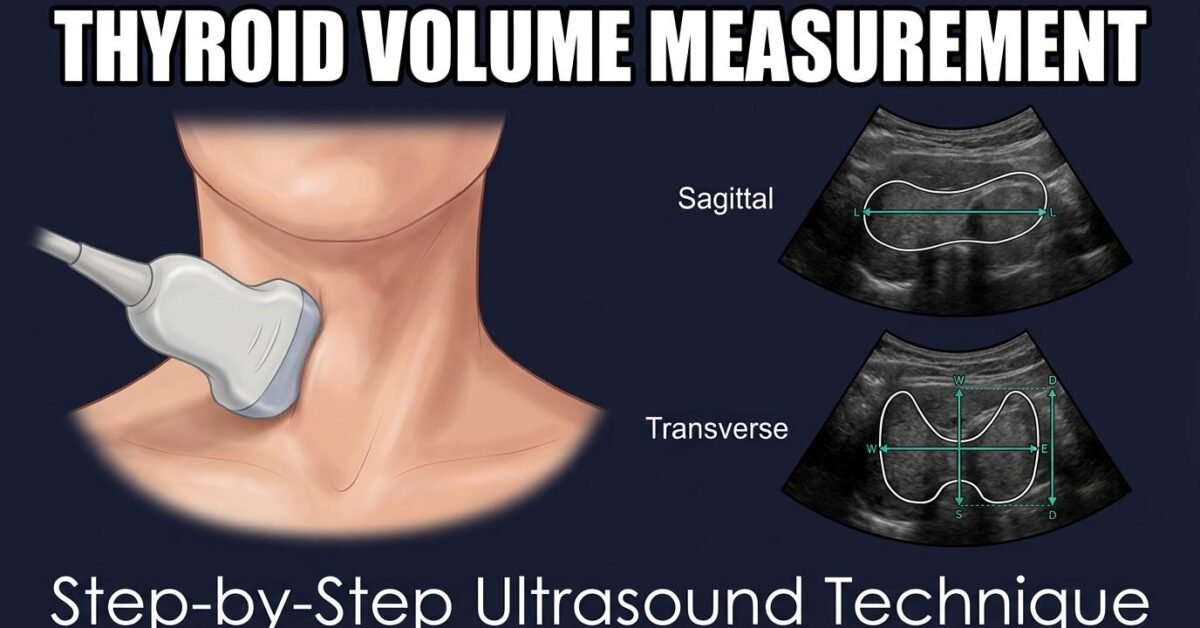
Accurate thyroid volume measurement is essential for diagnosing goiter, monitoring treatment response, and conducting population-based iodine surveys. This guide walks through the standard measurement technique, caliper placement, and common pitfalls.
Equipment and Patient Positioning
Use a high-frequency linear transducer (7 to 15 MHz) with the patient supine and the neck mildly hyperextended using a small pillow or rolled towel under the shoulders. Adequate neck extension improves access to the inferior poles, which are the most commonly undermeasured segments.
Step 1: Sagittal (Longitudinal) View
Position the transducer longitudinally along the long axis of each lobe. Identify the maximum craniocaudal extent of the lobe from the superior to inferior pole. Place electronic calipers at the extreme ends of the lobe to measure the length. A normal adult lobe measures approximately 40 to 60 mm.
Step 2: Transverse (Axial) View
Rotate the transducer 90 degrees at the mid-lobe level where the lobe appears widest. Measure the mediolateral dimension (width) from the lateral edge to the medial edge adjacent to the trachea. Measure the anteroposterior dimension (depth) from the anterior capsule to the posterior capsule. Exclude the strap muscles and carotid vessels from the measurement.
Step 3: Calculate the Volume
Apply the ellipsoid formula to each lobe separately:
Volume (mL) = Length (cm) × Width (cm) × Depth (cm) × 0.523
Sum the right and left lobe volumes for the total thyroid volume. The isthmus is excluded unless its AP thickness exceeds 3 mm, in which case its volume may be estimated and added. Use the thyroid volume calculator for quick computation.
Common Measurement Errors
The most frequent error is undermeasuring the length by not capturing the full extent of the inferior pole, especially in glands with retrosternal extension. Other common errors include measuring obliquely rather than in the true longitudinal or transverse plane, including perithyroidal structures such as strap muscle in the depth measurement, and inconsistent caliper placement between serial exams.
For clinicians: When monitoring volume over time, always measure in the same planes and use the same correction factor. A change of more than 20 percent between exams is considered clinically significant. If possible, have the same operator perform serial measurements to reduce inter-observer variability.
When to Use Alternative Methods
The ellipsoid method is adequate for most clinical purposes but underperforms in glands with markedly irregular morphology. For large multinodular goiters, retrosternal extension, or pre-surgical volumetry, consider 3D ultrasound volumetry or CT/MRI-based segmentation, which provide more accurate volume estimates.
Frequently Asked Questions
Which correction factor should I use?
Use 0.523 for routine clinical practice. Use 0.479 for WHO epidemiological comparison. Use 0.529 (Shabana) for maximum accuracy against CT volumetry. Whichever you choose, be consistent across serial exams.
Do I include the isthmus in the total volume?
Standard practice excludes the isthmus unless its AP thickness exceeds 3 mm.
How reproducible is thyroid volume measurement?
Intra-observer variability is typically 10 to 15 percent. Inter-observer variability can reach 20 to 25 percent. Consistent technique and operator are the most important factors.
