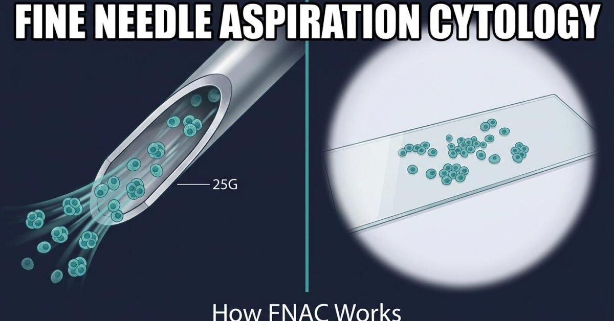
Fine needle aspiration cytology (FNAC) is the first-line sampling technique for thyroid nodules and many other superficial lesions. It uses a needle thinner than one used for routine venipuncture to collect individual cells for microscopic examination. This guide covers the technique, equipment, adequacy standards, and how FNAC differs from core needle biopsy.
What FNAC Actually Collects
The defining characteristic of FNAC is that it samples cells, not tissue. The aspirate contains individual cells and small cell clusters suspended in fluid, which are then spread onto slides. A pathologist evaluates the appearance of individual cells: nuclear size and shape, chromatin pattern, nuclear-to-cytoplasmic ratio, and the architectural groupings the cells form.
This is powerful for identifying malignancies with distinctive nuclear features, such as papillary thyroid carcinoma with its nuclear grooves and intranuclear pseudoinclusions. It is inherently limited for lesions where the diagnosis depends on architecture rather than cell appearance.
Equipment and Technique
Needle Selection
Thyroid FNA typically uses 25 to 27 gauge needles. The rationale for such fine needles is counterintuitive but well established: thinner needles collect less blood, and blood contamination is the single most common reason a slide becomes uninterpretable. A cleaner, less bloody sample yields better cytology even though fewer total cells are collected.
Aspiration vs Capillary Technique
Two approaches are used. In the aspiration technique, gentle negative pressure is applied with an attached syringe while the needle is moved within the lesion. In the capillary (non-aspiration or French) technique, no suction is applied and cells enter the needle purely by capillary action.
The capillary technique often produces less bloody samples and is favored for highly vascular lesions such as the thyroid. Many operators use both, reserving suction for fibrotic or sparsely cellular nodules.
Ultrasound Guidance
Real-time ultrasound guidance is standard for thyroid FNA and substantially reduces non-diagnostic rates compared with palpation-guided sampling. It allows the operator to confirm the needle tip is within the target, to sample the solid portion of partially cystic nodules, and to avoid vessels.
Number of Passes
Two to five passes are typical, sampling different regions of the nodule to account for heterogeneity. Each pass involves only a few seconds of needle movement within the lesion.
Sample Adequacy
For thyroid cytology, a sample is generally considered adequate when it contains at least six groups of well-preserved follicular cells, each group containing at least ten cells. Samples falling short are classified as Bethesda Category I (non-diagnostic).
Exceptions apply: a sample showing abundant colloid, or one with clear cytologic atypia, may be interpretable even without meeting the numeric threshold.
Rapid on-site evaluation (ROSE) by a cytotechnologist reduces non-diagnostic rates meaningfully, particularly for cystic and calcified nodules. Where ROSE is unavailable, increasing to four or five passes and targeting the solid mural component of complex cystic nodules achieves much of the same benefit.
FNAC vs Core Needle Biopsy
| FNAC | Core Needle Biopsy | |
|---|---|---|
| Sample type | Individual cells (cytology) | Tissue cylinder (histology) |
| Typical gauge | 25–27 G | 18–20 G |
| Architecture preserved | No | Yes |
| Discomfort | Minimal | Moderate |
| Bleeding risk | Very low | Low but higher |
| Anesthetic | Often none | Local required |
| Main limitation | Cannot assess capsular invasion | More invasive |
The practical consequence of the architecture difference is significant in the thyroid: FNAC cannot distinguish follicular adenoma from follicular carcinoma, because that distinction rests entirely on whether tumor cells have breached the capsule or invaded vessels. This is why Bethesda Category IV exists as a separate indeterminate category and why those cases often proceed to diagnostic lobectomy.
Applications Beyond the Thyroid
FNAC is used across many superficial and image-accessible sites, including cervical and axillary lymph nodes, salivary glands, breast lesions, and soft tissue masses. Lymph node FNA in the setting of suspected thyroid cancer is often paired with thyroglobulin measurement of the needle washout, which markedly increases sensitivity for metastatic disease.
Reporting
Thyroid FNAC results are reported using the Bethesda System, a six-category framework linking cytologic findings to malignancy risk and management. For the patient-facing view of the procedure, see our thyroid biopsy procedure guide.
Frequently Asked Questions
What is fine needle aspiration cytology?
Fine needle aspiration cytology (FNAC) is a diagnostic technique that uses a thin needle, typically 25 to 27 gauge, to collect individual cells from a lesion for microscopic examination. It samples cells rather than intact tissue, which distinguishes it from core needle biopsy.
What is the difference between FNA cytology and biopsy?
FNA cytology collects loose cells and evaluates their individual appearance. Core needle biopsy removes a small cylinder of tissue, preserving architecture so relationships between cells and structures such as a capsule can be assessed. FNAC is faster and less invasive; core biopsy provides architectural information FNAC cannot.
What needle gauge is used for thyroid FNA?
Thyroid FNA typically uses 25 to 27 gauge needles. Thinner needles reduce bleeding and blood contamination of the sample, which improves slide quality, and they cause less discomfort than larger-bore needles.
What makes an FNA sample adequate?
For thyroid cytology, adequacy generally requires at least six groups of well-preserved follicular cells with a minimum of ten cells per group. Samples not meeting this threshold are reported as Bethesda Category I, non-diagnostic.
