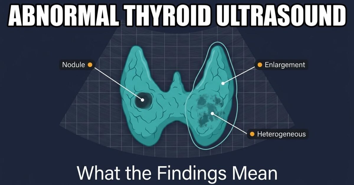
Being told your thyroid ultrasound is abnormal sounds alarming, but the word covers a wide range of findings, most of them benign and many requiring no treatment at all. This guide walks through the common abnormal findings and what each one actually means.
Thyroid Nodules
Nodules are by far the most common abnormal finding. They are present in up to 65 percent of adults when sensitive ultrasound is used, and prevalence rises with age. The overwhelming majority are benign.
Each nodule is scored across five ultrasound features and assigned a TI-RADS category from TR1 to TR5. The category, combined with the nodule’s size, determines whether biopsy, surveillance, or no action is recommended:
| Category | Malignancy risk | Biopsy threshold |
|---|---|---|
| TR1 / TR2 | 0.3% / 1.5% | No biopsy at any size |
| TR3 | ~4.8% | ≥ 2.5 cm |
| TR4 | ~9.1% | ≥ 1.5 cm |
| TR5 | ~35% | ≥ 1.0 cm |
Score yours with the TI-RADS calculator.
Heterogeneous Thyroid Gland
A heterogeneous thyroid has an uneven, coarsened texture instead of the normal fine and uniform appearance. This describes the whole gland rather than a discrete nodule.
The most common cause by a wide margin is chronic autoimmune thyroiditis, particularly Hashimoto disease, where lymphocytic infiltration disrupts the normal tissue architecture. Other causes include multinodular change and, less often, subacute thyroiditis.
A heterogeneous gland is a description, not a diagnosis. It is interpreted alongside thyroid function tests and antibody levels. Many people with heterogeneous glands have entirely normal thyroid function and need no treatment.
Enlarged Thyroid
Total gland volume above the reference range indicates enlargement, described clinically as goiter. Reference upper limits are approximately 15 mL in women and 18 mL in men.
Causes range from iodine deficiency, the leading cause worldwide, to autoimmune conditions and multinodular change. Read more in our guide to enlarged thyroid volume, or measure your own gland with the thyroid volume calculator.
Small or Atrophic Thyroid
A gland below the normal size range may reflect long-standing autoimmune thyroiditis where functioning tissue has been progressively replaced by fibrosis, or prior treatment with radioactive iodine or surgery. Thyroid function tests usually accompany this finding.
Increased Vascularity
Colour Doppler showing markedly increased blood flow throughout the gland, sometimes called a thyroid inferno, is characteristic of Graves disease. This is a functional diagnosis confirmed with blood tests and sometimes an uptake scan rather than by imaging alone.
Abnormal Lymph Nodes
Normal cervical lymph nodes are oval with a bright fatty hilum. Features that raise concern include a rounded shape, loss of the hilum, internal microcalcifications, cystic change, or chaotic peripheral vascularity.
Suspicious nodes in a patient with a thyroid nodule are significant and may warrant sampling of the node itself, often with thyroglobulin measurement of the needle washout.
Abnormal on a thyroid ultrasound rarely means dangerous. Ask your doctor which specific finding was abnormal, whether it affects thyroid function, and whether anything needs to be done or simply monitored.
Distinguish clearly in the report between diffuse parenchymal change and discrete nodules, since the two drive different pathways. Heterogeneity alone does not warrant FNA; apply TI-RADS to discrete nodules only.
What Happens Next
Depending on the finding, the outcome is typically one of:
- No action — benign-appearing findings below action thresholds
- Blood tests — TSH, free T4, and thyroid antibodies to assess function
- Surveillance ultrasound — repeat imaging at intervals defined by the TI-RADS category
- Fine needle aspiration — where a nodule meets the size and category threshold
To read your report term by term, see understanding your thyroid ultrasound report, or the full thyroid ultrasound guide.
Frequently Asked Questions
What does an abnormal thyroid ultrasound mean?
An abnormal result simply means something differs from the expected appearance. The most common findings are nodules, gland enlargement, or a heterogeneous texture. The great majority of abnormal thyroid ultrasounds reflect benign conditions.
What does heterogeneous thyroid mean?
A heterogeneous thyroid has an uneven, patchy texture rather than the normal uniform appearance. It is most commonly caused by chronic autoimmune thyroiditis such as Hashimoto disease, and is a description of the gland rather than a diagnosis.
Is an abnormal thyroid ultrasound serious?
Usually not. Thyroid nodules are found in up to 65 percent of adults and the vast majority are benign. Seriousness depends on the specific finding, its TI-RADS category if a nodule is present, and your thyroid function tests.
What happens after an abnormal thyroid ultrasound?
Next steps depend on the finding. Options include no action, repeat ultrasound for surveillance, thyroid function blood tests, or fine needle aspiration biopsy if a nodule meets the size and category threshold.
