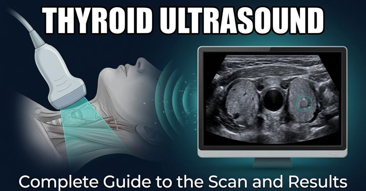
A thyroid ultrasound is the primary imaging test for evaluating the thyroid gland. It uses high-frequency sound waves to produce real-time images of the gland’s size, texture, and any nodules within it, without radiation and without discomfort. This guide covers why the scan is ordered, what happens during it, and how to interpret the report you receive afterward.
Why a Thyroid Ultrasound Is Ordered
Your doctor may request a thyroid ultrasound for several reasons:
- A lump you or your doctor can feel in the front of the neck
- An incidental finding on a CT, MRI, or PET scan performed for another reason
- Abnormal thyroid function tests, to look for structural causes
- Known nodules requiring surveillance to track size or feature changes
- Suspected goiter or a visibly enlarged gland
- Neck lymph node assessment, particularly where thyroid cancer is suspected or being followed
- Guidance during a biopsy, to direct the needle in real time
What a Thyroid Ultrasound Shows
Ultrasound is excellent at characterizing structure. A thyroid scan reports on:
- Gland size — three dimensions per lobe, converted to a volume in millilitres
- Echotexture — whether the tissue looks uniform or coarsened and heterogeneous
- Nodules — their number, size, location, and detailed features
- Blood flow — assessed with Doppler imaging
- Lymph nodes — size, shape, and internal architecture of neck nodes
What it cannot show is function. An ultrasound reveals nothing about whether your thyroid is producing too much or too little hormone; that requires blood tests. It also cannot definitively diagnose cancer, only estimate risk.
How to Prepare
Preparation is minimal. You do not need to fast or stop any medication. Wear a shirt with an open or low neckline, and leave necklaces at home or remove them before the scan. Allow around 30 minutes for the appointment.
What Happens During the Scan
- Positioning. You lie on your back with a pillow under your shoulders so the neck is gently extended, bringing the thyroid closer to the surface.
- Gel application. Warm water-based gel is spread over the front of the neck. It eliminates air between the probe and skin so sound waves transmit cleanly.
- Scanning. The sonographer moves a small handheld probe over the neck in both transverse and sagittal planes, capturing images of each lobe, the isthmus, and surrounding structures.
- Measurements. Electronic calipers are placed to measure the gland and any nodules found.
- Doppler. Colour Doppler may be applied to assess blood flow within nodules or the gland.
- Finishing. The gel is wiped off and you can leave immediately. There are no after-effects and no restrictions.
The scan is painless. You may feel mild pressure from the probe and be asked to swallow or tilt your head at points.
Understanding Thyroid Ultrasound Images
Ultrasound images are composed of shades of gray representing how strongly different tissues reflect sound. Normal thyroid tissue appears as a uniform, moderately bright (slightly hyperechoic) structure sitting either side of the trachea, with the carotid arteries visible laterally as dark round channels.
Terms describing brightness relative to that normal tissue appear throughout reports:
| Term | Appearance | Typical meaning |
|---|---|---|
| Anechoic | Black | Pure fluid, simple cyst |
| Hypoechoic | Darker than thyroid | Solid tissue |
| Isoechoic | Same as thyroid | Often benign nodule |
| Hyperechoic | Brighter than thyroid | Fat, calcium, fibrous tissue |
Our echogenicity guide covers these terms in depth, and the TI-RADS examples page describes what each ultrasound feature looks like.
Normal Thyroid Ultrasound Findings
A normal thyroid gland shows a homogeneous, fine echotexture with smooth capsular margins. Reference dimensions for adults:
| Measurement | Normal range |
|---|---|
| Lobe length | 4.0 – 6.0 cm |
| Lobe width | 1.3 – 1.8 cm |
| Lobe depth | 1.3 – 1.8 cm |
| Isthmus thickness | < 0.3 cm |
| Total volume (female) | Up to 15 mL |
| Total volume (male) | Up to 18 mL |
Calculate your own with the thyroid volume calculator, or read the full reference ranges in our normal thyroid size guide.
Abnormal Thyroid Ultrasound Findings
Common abnormal findings include:
- Nodules — extremely common, found in up to 65 percent of adults on sensitive scanning. The vast majority are benign.
- Diffuse enlargement (goiter) — total volume above the gender-specific reference range
- Heterogeneous echotexture — a coarsened, patchy appearance often seen in Hashimoto thyroiditis
- Increased vascularity — a hypervascular gland on Doppler can suggest Graves disease
- Abnormal lymph nodes — rounded shape, loss of the fatty hilum, or internal calcification
Thyroid Nodules on Ultrasound and TI-RADS
When a nodule is found, the radiologist scores five ultrasound features to estimate its risk of malignancy: composition, echogenicity, shape, margin, and echogenic foci. The points are added and the total assigns a category from TR1 to TR5 under the ACR TI-RADS system.
| Category | Points | Malignancy risk | Biopsy threshold |
|---|---|---|---|
| TR1 | 0 | ~0.3% | None |
| TR2 | 2 | ~1.5% | None |
| TR3 | 3 | ~4.8% | ≥ 2.5 cm |
| TR4 | 4–6 | ~9.1% | ≥ 1.5 cm |
| TR5 | 7+ | ~35% | ≥ 1.0 cm |
Enter your nodule’s features into the TI-RADS calculator to see the category and recommendation, or view every point value on the TI-RADS chart.
Finding a thyroid nodule on ultrasound is common and usually not a cause for alarm. Ask your doctor for two things: the TI-RADS category and the nodule’s size in centimetres. Those two together determine whether anything needs to be done.
Scan in both transverse and sagittal planes and image the inferior poles fully, as undermeasurement there is the commonest source of volume error. Assess cervical node compartments in any patient with a TR4 or TR5 nodule, since nodal status changes management independently of the primary nodule’s size.
Thyroid Ultrasound vs Thyroid Scan
These two tests are frequently confused but answer different questions:
| Thyroid ultrasound | Thyroid uptake scan | |
|---|---|---|
| Technology | Sound waves | Radioactive tracer |
| Shows | Structure and anatomy | Function and hormone activity |
| Radiation | None | Low dose |
| Used for | Nodules, size, biopsy guidance | Hyperthyroidism, hot vs cold nodules |
Read more in our thyroid uptake scan guide.
What Happens After the Scan
A radiologist reviews the images and issues a report, usually within a few days. Depending on the findings, the outcome may be no further action, scheduled surveillance imaging, or a recommendation for fine needle aspiration biopsy.
To interpret your report line by line, see understanding your thyroid ultrasound report. If biopsy is recommended, our thyroid biopsy guide explains what to expect.
Frequently Asked Questions
What is a thyroid ultrasound?
A thyroid ultrasound is a painless imaging test that uses high-frequency sound waves to create real-time pictures of the thyroid gland in the neck. It shows the gland’s size, texture, and any nodules, and it involves no radiation.
Do I need to prepare for a thyroid ultrasound?
No special preparation is needed. You do not need to fast, and you can take your usual medications. Wear a top with an open neckline and remove necklaces before the scan.
How long does a thyroid ultrasound take?
A thyroid ultrasound typically takes 15 to 30 minutes. If neck lymph nodes are also examined, it may take slightly longer.
Can a thyroid ultrasound detect cancer?
An ultrasound cannot diagnose cancer on its own. It identifies features that suggest higher or lower risk, which are scored using systems such as ACR TI-RADS. Only a biopsy can confirm whether a nodule is cancerous.
Does a thyroid ultrasound show thyroid function?
No. Ultrasound shows the gland’s structure and appearance, not how well it is working. Thyroid function is assessed with blood tests measuring TSH, T4, and T3.
