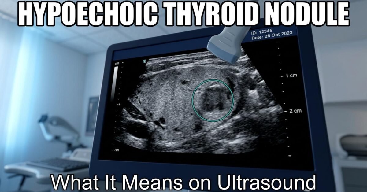
If your ultrasound report describes a hypoechoic nodule, a hypoechoic lesion, or a hypoechoic mass, it means the structure appeared darker than the tissue around it. Echogenicity is one of five features scored in the ACR TI-RADS system, and how dark a nodule appears helps estimate its risk. This guide covers the full echogenicity scale, what each term means, how many TI-RADS points each earns, and what happens next.
Hypoechoic Definition
Hypoechoic means darker than the surrounding tissue on an ultrasound image. The prefix “hypo” means less, and “echoic” refers to the echoes that create the ultrasound picture. A hypoechoic structure reflects fewer sound waves back to the probe, so it appears darker on screen.
On a thyroid ultrasound specifically, hypoechoic means the nodule looks darker than the normal thyroid tissue surrounding it. The same term is used throughout the body, so a hypoechoic lesion in the liver, breast, or kidney carries the same basic meaning: darker than its neighbouring tissue.
You may also see the related terms hypoechogenicity (the property of being hypoechoic) or hypoechogenic (an alternative adjective form). All three describe the same finding.
The Complete Echogenicity Scale
Ultrasound images are made of shades of gray. Radiologists use a standard vocabulary to describe where a structure sits on that brightness scale:
| Term | Appearance | Compared to | Typical cause | TI-RADS points |
|---|---|---|---|---|
| Anechoic | Pure black | No echoes at all | Simple fluid, cysts | 0 |
| Hypoechoic | Darker gray | Darker than thyroid tissue | Solid tissue, some cancers | 2 |
| Isoechoic | Medium gray | Same as thyroid tissue | Benign nodules | 1 |
| Hyperechoic | Bright / whiter | Brighter than thyroid tissue | Fat, calcium, fibrous tissue | 1 |
| Very hypoechoic | Near black | Darker than neck muscle | Most concerning finding | 3 |
Enter your nodule’s echogenicity along with its other features into the TI-RADS calculator to see the total score and category.
Hyperechoic vs Hypoechoic
These two terms sit on opposite sides of the echogenicity scale and are frequently confused.
| Hypoechoic | Hyperechoic | |
|---|---|---|
| Appearance | Darker than surrounding tissue | Brighter than surrounding tissue |
| Sound reflection | Reflects fewer echoes | Reflects more echoes |
| Common causes | Solid cellular tissue, some malignancies, inflammation | Fat, calcification, fibrous tissue, colloid |
| TI-RADS points | 2 points | 1 point |
| Relative concern | Higher | Lower |
In practical terms, hyperechoic nodules are more often benign, which is why they score only 1 point compared to 2 for hypoechoic. However, echogenicity alone never determines whether a nodule is cancerous.
Isoechoic: The Middle Ground
Isoechoic means the structure has the same brightness as the surrounding tissue. An isoechoic nodule blends into the thyroid because it reflects sound waves at roughly the same rate as normal thyroid tissue.
This can make isoechoic nodules harder to detect. They are often identified by a subtle halo around them, a change in the gland’s contour, or displacement of nearby structures rather than by a difference in brightness.
Isoechoic vs Hypoechoic
The difference is straightforward: an isoechoic nodule matches the thyroid’s brightness, while a hypoechoic nodule is visibly darker than it. In ACR TI-RADS, isoechoic scores 1 point (grouped with hyperechoic), while hypoechoic scores 2 points.
Isoechoic Nodules in the Thyroid
An isoechoic thyroid nodule is generally a reassuring finding on the echogenicity axis. Most isoechoic nodules are benign, commonly representing hyperplastic or adenomatous nodules. A solid isoechoic thyroid nodule still earns 2 points for its solid composition plus 1 point for echogenicity, so the combination matters more than either feature alone.
You may also encounter the term isoechogenic, which means the same thing as isoechoic.
Anechoic vs Hypoechoic
Anechoic means completely black with no internal echoes whatsoever. Hypoechoic means dark gray, but with some internal echoes present.
The distinction matters clinically because anechoic almost always indicates pure fluid, such as a simple cyst, which carries essentially no cancer risk and scores 0 TI-RADS points. Hypoechoic indicates solid tissue that is darker than its surroundings, which is why it scores 2 points.
A hypoechoic cyst is a slightly different finding. A truly simple cyst should be anechoic. When a cyst appears hypoechoic rather than black, it usually contains debris, blood products, or proteinaceous material rather than clear fluid. This is often still benign but may be described as a complex cyst.
Heterogeneous Echogenicity
Heterogeneous means the echogenicity is not uniform throughout the structure. A heterogeneous hypoechoic mass is predominantly darker than surrounding tissue but contains areas of varying brightness within it, giving it a mottled or patchy internal appearance.
Heterogeneous echogenicity can result from several processes, including areas of hemorrhage, necrosis, cystic degeneration, calcification, or fibrosis within a nodule. It is a descriptive term rather than a diagnosis, and it appears in both benign and malignant nodules.
In the thyroid gland as a whole, a heterogeneous appearance across the entire gland (rather than within one nodule) is a common finding in chronic autoimmune conditions such as Hashimoto thyroiditis.
Heterogeneous Hypoechoic Mass
When a report describes a heterogeneously hypoechoic mass, it means the lesion is mostly dark with internal variation. This combination warrants attention because both features, low echogenicity and internal heterogeneity, appear more frequently in malignant lesions than in simple benign ones. The nodule’s full TI-RADS score, including margin, shape, and echogenic foci, determines the actual risk level.
Hypoechoic Lesions Elsewhere in the Body
The terms hypoechoic lesion, hypoechoic mass, hypoechoic area, hypoechoic focus, and hypoechoic structure all describe the same fundamental finding in different contexts. The clinical meaning depends entirely on which organ is being scanned:
- Thyroid — scored within the TI-RADS system, as described throughout this article
- Breast — assessed using BI-RADS; hypoechoic is a standard descriptor for solid masses
- Liver — may represent hemangioma, focal nodular hyperplasia, metastasis, or other lesions
- Kidney — evaluated for solid versus cystic characteristics
- Lymph nodes — normal nodes have a hypoechoic cortex with an echogenic hilum
A hypoechoic avascular mass means the lesion shows no blood flow on Doppler imaging, which in some contexts favors a benign process, though this must be interpreted alongside all other findings.
Why Hypoechoic Matters in TI-RADS
Studies have consistently linked low echogenicity to a higher likelihood of thyroid malignancy, particularly papillary thyroid carcinoma. This is why the ACR TI-RADS system assigns 2 points to hypoechoic nodules and 3 points to very hypoechoic nodules.
That said, most hypoechoic thyroid nodules are benign. Hypoechogenicity is one of the most common descriptions in thyroid ultrasound reporting. The finding earns points, but the final category depends on all five features combined.
Hypoechoic vs Very Hypoechoic
The distinction rests on the comparison reference:
- Hypoechoic (2 points) — darker than the normal thyroid parenchyma
- Very hypoechoic (3 points) — darker than the anterior strap muscles of the neck
Very hypoechoic is the more concerning of the two and carries the strongest association with malignancy of any echogenicity finding.
Reserve the “very hypoechoic” designation for nodules genuinely darker than the adjacent strap musculature. Overusing it inflates TR category and increases unnecessary biopsy rates. When echogenicity truly cannot be determined, ACR TI-RADS assigns 1 point rather than defaulting to a higher-risk category.
Seeing “hypoechoic” on your report is extremely common and is not alarming on its own. Ask your doctor for two specific pieces of information: the full TI-RADS category and the nodule’s size in centimetres. Those two together determine whether anything needs to be done.
How Echogenicity Combines With Other Features
Echogenicity is one of five scored features. Here is how the same hypoechoic finding produces very different outcomes depending on what accompanies it:
| Scenario | Features | Total | Category |
|---|---|---|---|
| Hypoechoic, otherwise reassuring | Solid (2) + hypoechoic (2) + wider-than-tall (0) + smooth (0) + no foci (0) | 4 | TR4 |
| Hypoechoic with suspicious features | Solid (2) + hypoechoic (2) + taller-than-wide (3) + irregular (2) + punctate foci (3) | 12 | TR5 |
| Mixed with hypoechoic component | Mixed (1) + hypoechoic (2) + wider-than-tall (0) + smooth (0) + none (0) | 3 | TR3 |
See the complete point breakdown on the TI-RADS chart, or view what each feature looks like on ultrasound in our TI-RADS examples guide.
What Happens Next
Whether a hypoechoic nodule requires biopsy depends on its total TI-RADS category and its size, not on echogenicity alone. General thresholds are:
- TR3 — FNA at 2.5 cm or larger
- TR4 — FNA at 1.5 cm or larger
- TR5 — FNA at 1.0 cm or larger
Smaller nodules in these categories are typically monitored with follow-up ultrasound. Read more in our guides to TI-RADS 4 and TI-RADS 5.
Frequently Asked Questions
What does hypoechoic mean?
Hypoechoic means darker than the surrounding tissue on an ultrasound image. The structure reflects fewer sound waves back to the probe, producing a darker appearance on screen. In the thyroid it earns 2 TI-RADS points.
What is the difference between hyperechoic and hypoechoic?
Hypoechoic is darker than the surrounding tissue and scores 2 TI-RADS points. Hyperechoic is brighter than the surrounding tissue and scores 1 point. Hyperechoic findings often reflect fat, calcification, or fibrous tissue and are generally less concerning.
What does isoechoic mean?
Isoechoic means the structure has the same brightness as the surrounding tissue, so it blends in rather than standing out. Isoechoic nodules score 1 TI-RADS point and are most often benign.
What is the difference between anechoic and hypoechoic?
Anechoic means completely black with no internal echoes, which indicates pure fluid such as a simple cyst and scores 0 points. Hypoechoic means dark gray with some internal echoes, indicating solid tissue, and scores 2 points.
What does heterogeneous hypoechoic mean?
It means the lesion is predominantly darker than surrounding tissue but has an uneven, patchy internal appearance rather than a uniform one. This can result from hemorrhage, necrosis, cystic change, or calcification within the nodule.
Is a hypoechoic nodule cancer?
Usually not. Hypoechogenicity is one of the most common findings in thyroid ultrasound and the majority of hypoechoic nodules are benign. It contributes 2 points toward the TI-RADS score, but the overall category and nodule size determine whether biopsy is needed.
What is a hypoechoic mass?
A hypoechoic mass is any solid lesion that appears darker than the tissue around it on ultrasound. The clinical significance depends on the organ involved, the mass’s other imaging features, and its size.
What does hypoechogenicity mean?
Hypoechogenicity is the property of being hypoechoic, meaning reduced echo reflection compared with surrounding tissue. It is the noun form of the same finding described by the adjective hypoechoic.
What is the difference between hypoechoic and very hypoechoic?
Hypoechoic means darker than the surrounding thyroid tissue and scores 2 TI-RADS points. Very hypoechoic means darker than the neck strap muscles, which is a more concerning finding and scores 3 points. The comparison reference changes between the two.
Is a hypoechoic nodule always dangerous?
No. Hypoechoic is one of the most common descriptions for thyroid nodules, and the majority of hypoechoic nodules are benign. It scores 2 points in TI-RADS but the overall category depends on all five features combined, not echogenicity alone.
Do I need to worry if my thyroid ultrasound shows punctate echogenic foci?
Punctate echogenic foci score 3 points in TI-RADS and are the most concerning type of echogenic foci because they may represent microcalcifications associated with papillary thyroid cancer. However, they can also occur in benign nodules. The overall TI-RADS category and nodule size determine whether action is needed.
Is it normal to have a thyroid nodule with calcifications?
Calcifications in thyroid nodules are not uncommon. Macrocalcifications (large calcium deposits) score 1 TI-RADS point and are seen in both benign and malignant nodules. Peripheral rim calcifications score 2 points. Punctate echogenic foci (microcalcifications) score 3 points and carry more concern. Having calcifications does not automatically mean cancer.
