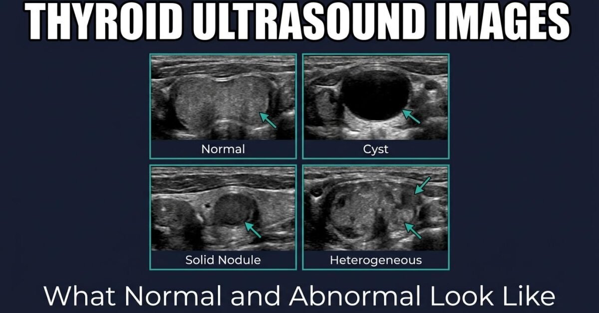
Thyroid ultrasound images are made of shades of gray, and learning to read them starts with knowing what normal looks like. This guide describes the appearance of a healthy gland and the common abnormal findings, so you can follow along with the descriptions in your report.
How Ultrasound Images Are Formed
The probe emits high-frequency sound waves that travel into the neck and bounce back at tissue boundaries. Strong reflectors appear bright; weak reflectors appear dark. Fluid transmits sound almost completely and so appears black. This is why the vocabulary of thyroid reporting is entirely about relative brightness.
Normal Thyroid Ultrasound Appearance
On a transverse image through the mid-neck, a normal thyroid is recognisable by a consistent set of landmarks:
- Two lobes sitting either side of the trachea, which appears as a bright curved line with dark shadowing behind it
- The isthmus crossing the front of the trachea as a thin band, normally under 3 mm thick
- Uniform echotexture — fine, even, and slightly brighter than the strap muscles lying in front of the gland
- Carotid arteries visible laterally as round black channels, pulsatile in real time
- Smooth capsular margins with clear separation from surrounding tissue
Reference dimensions and volumes are in our normal thyroid size guide.
Common Findings on Thyroid Ultrasound Images
Simple Cyst
A round, completely black (anechoic) structure with thin smooth walls. Sound passes through fluid unimpeded, producing a bright band behind the cyst known as posterior acoustic enhancement. Simple cysts are essentially never malignant and score 0 TI-RADS points.
Spongiform Nodule
Multiple tiny cystic spaces packed together produce a honeycomb or sponge-like appearance. When more than half the nodule shows this pattern, it is considered a benign finding and scores 0 points.
Solid Nodule
A discrete area of uniform tissue with no fluid spaces. Solid composition scores 2 points because virtually all thyroid cancers are solid, though the great majority of solid nodules are still benign.
Mixed Cystic and Solid Nodule
Both fluid and tissue components within one nodule. The solid portion is the part that matters for risk assessment and is where a biopsy needle should be directed.
Calcifications
Bright spots within a nodule. Large macrocalcifications cast a dark acoustic shadow behind them. Peripheral calcification forms a bright rim around the nodule edge. Tiny non-shadowing punctate foci are the most concerning type and score 3 points.
Heterogeneous Gland
A coarsened, patchy texture across the whole gland rather than within a single nodule. This is characteristic of chronic autoimmune thyroiditis such as Hashimoto disease.
Features That Raise Concern
Four appearances shift a nodule toward higher TI-RADS categories:
| Feature | What it looks like | Points |
|---|---|---|
| Very hypoechoic | Darker than the neck strap muscles | 3 |
| Taller-than-wide | Vertical orientation on transverse view | 3 |
| Irregular margin | Jagged, spiculated, or lobulated edges | 2 |
| Punctate echogenic foci | Tiny bright dots without shadowing | 3 |
See each feature described in detail on our TI-RADS examples page.
Looking at your own ultrasound images rarely helps and often causes unnecessary worry, because appearance alone does not determine risk. The TI-RADS category and nodule size in your report are the meaningful numbers.
Doppler Imaging
Colour Doppler overlays blood flow onto the grayscale image. Peripheral flow around a nodule’s rim is common in benign lesions, while chaotic internal vascularity is seen more often in malignancy. Doppler findings are supportive rather than decisive and are not part of the ACR TI-RADS point system.
From Image to Decision
What matters is not any single image but the combined score. Enter the features described in your report into the TI-RADS calculator to see the category, malignancy risk, and whether biopsy is indicated. For the full scan overview, see our thyroid ultrasound guide.
Frequently Asked Questions
What does a normal thyroid look like on ultrasound?
A normal thyroid appears as a uniform, moderately bright structure on either side of the trachea, slightly brighter than the adjacent strap muscles. The texture is fine and homogeneous, and the capsule edges are smooth and well defined.
What does a thyroid nodule look like on ultrasound?
A nodule appears as a discrete area that differs from surrounding thyroid tissue. It may be darker (hypoechoic), the same brightness (isoechoic), brighter (hyperechoic), or black if fluid-filled. Its margins, shape, and any bright internal spots are all assessed.
Can you tell if a thyroid nodule is cancerous from the ultrasound image?
Not definitively. Ultrasound identifies features associated with higher or lower risk, which are scored using TI-RADS. Even the highest category, TR5, is benign about two thirds of the time. Only biopsy provides a diagnosis.
What does Hashimoto thyroiditis look like on ultrasound?
Hashimoto thyroiditis typically produces a heterogeneous, coarsened echotexture throughout the gland, often with a hypoechoic background and fine echogenic septations. The gland may be enlarged early in the disease and atrophic later.
